


CT scan
You will need a CT scan before treatment to show the location and size of the liver tumours. CT scans you have had recently may be sufficient for measuring the liver tumour, but sometimes they have to be repeated.
Work-up procedure
You will need to attend a pre-treatment day when you will need to have some tests to help prepare you for treatment (called the ‘work-up’ procedure). You will then be invited back usually one-to-two weeks later, for treatment using the SIRT procedure. To assess your suitability for the treatment, the following tests will be performed during the work-up:
1. Liver angiogram
2. Lung-shunting scan
After these tests, you can usually go home on the same day. Occasionally, these tests reveal that treatment using SIRT is not achievable because the microspheres cannot be safely delivered to liver tumours without damaging other sensitive tissue.
Based on the results of these tests, it will be decided whether you can have SIRT and the dose will be ordered so that it is ready for the day of your treatment.
1) Liver angiogram
The liver angiogram provides a detailed picture of the blood supply to the liver, which can vary between people. The aim of this procedure is to look at the blood supply to the tumour and to block off (‘embolise’) tiny blood vessels that lead to healthy organs of the body, such as the lungs or the stomach. This will stop the microspheres from damaging healthy tissue when they are administered to you on the treatment day, ensuring that they are only delivered to the tumour.
The blood vessels in the liver are a bit like a tree with lots of branches. With this in mind, you can think about this procedure as your doctor ‘pruning’ back some of the branches of the tree to ensure that the treatment goes directly to where it’s needed.
Before the angiogram, the groin area is numbed with a local anaesthetic and a small cut is made. Some sedation may also be used to make you feel comfortable. A soft, flexible tube (catheter) is inserted through the cut and guided through a blood vessel (femoral artery) into the liver using x-ray pictures.
A dye, called a contrast medium, will be injected through the catheter to show up the blood vessels in the liver. The doctor then blocks off the vessels that lead to healthy parts of the body.
The procedure usually takes about 60-90 minutes but may take longer in some cases.
It usually involves little discomfort and you may have a feeling of warmth or a slight burning sensation when the contrast material is injected. The most difficult part may be lying flat for the procedure. After the procedure, you can resume a normal diet and all normal activities within eight to ten hours.
There is no risk associated with blocking off these tiny blood vessels, but it can sometimes cause mild pain for a few hours after the procedure, so you will be monitored in the hospital for at least four hours before you can go home.
On the same day, this procedure will include a scan of your liver to calculate the correct dose of treatment that will be required on your treatment day.
Sometimes the information given in the liver angiogram is not sufficient. Your doctor might decide to do an additional test called a CT scan. A dye called a contrast medium is injected through a catheter in your groin, while the scan is performed to show the blood vessels in the liver.
2) Lung-shunting scan
This test is also known as a scintigraphy (sin-tig-raf-ee) or a MAA scan. It involves injecting a radioactive dye into the catheter that has already been inserted during the liver angiogram procedure. The dye mimics what the microspheres will do on the day of treatment and allows your doctor to predict where they will lodge themselves on the day of treatment.
Sometimes some of the dye will pass through the liver into the lung. Your doctor needs to know how much passes through to determine a safe dose of microspheres for you. You will need to remain still for the brief periods of time during this procedure while scans are taken.
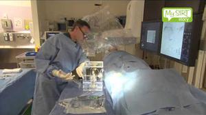
Watch the patient procedure video below:
There are three ways in which you can get SIRT treatment: