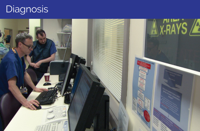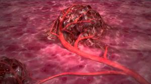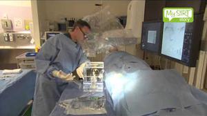
Liver tumours can be diagnosed using a combination of tests. They will often show up on an ultrasound scan, but full assessment requires a CT scan and/or MRI scan. If the results from blood tests and CT or MRI scans are not obvious, a biopsy may be required to confirm the diagnosis, where a small piece of tissue from the affected area is removed and assessed. The different types of tests that may be used are described in the following sections.
Physical exam and history
Your doctor will examine your body to check your general health, including any signs of disease (such as lumps or weight loss) or anything else that seems unusual. The doctor will also take a history of your health including past illnesses (especially hepatitis) and will ask about any symptoms you are experiencing.
Blood tests
Liver function tests (LFT)
These blood tests show if the liver is working properly. It is important to realise that normal liver function can be affected by many conditions other than cancer. These tests are also useful to see how well the liver works before, during and after treatment.
Serum tumour marker test
These are blood tests that look for certain substances called tumour markers. If these are present in higher than usual levels then it can mean that there are active cancer cells in the body.
AFP (Alpha-fetoprotein) in the blood may be a sign of primary liver cancer. Other cancers and some non-cancerous conditions, such as cirrhosis (scarring of the liver) and hepatitis (liver inflammation), may also increase AFP levels.
CEA (carcinoembryonic antigen) is very high in patients with liver cancer that has spread from the bowel. It may also be high in people with other cancers of the gut, lung, and breast as well as in some non-cancerous diseases and healthy smokers.
Scans
Ultrasound scan
This test uses sound waves that bounce off your internal organs and tissues to create a picture of a part of the body. The scan will show any abnormal growths in your liver. You may be asked not to eat or drink for at least four hours before the scan. The scan takes only a few minutes and is painless. You usually sit or lie near the ultrasound machine. A clear gel is spread on your skin over the area that will be scanned. The gel helps to transmit the sound waves. You can usually go home as soon as the scan is over. CT (computerised tomography) scan
A CT scan takes many x-rays to create a very detailed three-dimensional picture of the inside of the body. This type of scan may be used to look for signs of cancer. A CT scan can give a very accurate picture of the location and size of a tumour. It can also show how close major body organs are to the area that needs to be treated or operated on. A dye called a contrast medium may be injected partway through the scan to help show up more detail of your liver and the tumours. A CT scanning machine is large and "doughnut" shaped. You lie on a bed that can slide backwards and forwards through the hole of the machine. The pictures are taken as you move through the machine. An abdominal (tummy) CT scan takes less than 10 minutes and is painless. You can usually go home as soon as the scan is over. CT is also called computed axial tomography (CAT).
MRI (magnetic resonance imaging) scan
An MRI scan is similar to a CT scan but uses magnetic fields instead of x-rays to build up a picture of inside the body. An MRI scan will be clearer than a CT scan for some types of tissues. Your doctor will know the best scan for you. The MRI scanner is a large cylinder with a bed that can move backwards and forwards through the cylinder. You will need to remove all metal belongings before going into the room because the scanner is a very powerful magnet. You may have an injection of a dye called a contrast medium just before the scan. This dye helps to show body tissues and organs more clearly on the scan. MRI is painless, but the machine can be noisy. You may be given earplugs or headphones to wear. The test is likely to take less than half an hour, but may be up to an hour. You are usually able to go home as soon as the scan is over. MRI is also called nuclear magnetic resonance imaging (NMRI).
PET (positron emission tomography) scan
A PET scan uses an injected mildly radioactive sugar that shows up where cancer cells are active in the body. A CT scan is then performed in a machine called a PET-CT to show where these radioactive ‘cancer hot-spots’ are located.
Biopsy
One way to diagnose cancer is to take a sample of the tumour. This is called a biopsy. The sample is viewed under a microscope. It can show whether the cancer is primary or secondary liver cancer.
You may have a biopsy at the same time as an ultrasound or CT scan, usually through a needle. The skin is numbed with a local anaesthetic and the needle is inserted into the liver through the skin.
A biopsy is usually only done if other tests including blood tests and CT or MRI scan do not have obvious results. A biopsy carries a small risk of bleeding and that the tumour will spread along the needle track, so is not always thought to be necessary by doctors.
Following a liver biopsy, you may have to stay in hospital for a few hours or overnight. This is because the liver has a very rich blood supply and there is a risk of bleeding afterwards.
Laparoscopy
This involves a small operation that allows a doctor access to the abdomen (tummy) using only a small incision in the skin. This is sometimes referred to as keyhole surgery. A thin fibreoptic telescope with a camera attached (laparoscope) is passed through the hole into the liver where the liver can be examined and a small piece of tissue removed and examined under a microscope (biopsy). A small cut in the abdomen will be made under anaesthetic so the laparoscope can be inserted into the liver. A few stitches will be required afterwards and you may have to stay in hospital for a day or so.
Liver angiogram
This allows doctors to look at the blood supply to the liver and how the tumour may be affecting it. A fine tube is inserted into an artery in your groin and a dye is injected through the tube. The dye circulates around your blood vessels and is shown up using an x-ray. More details can be found in the before treatment section.

There are three ways in which you can get SIRT treatment:


